Echocardiogram (Heart Ultrasound) in Birmingham: What It Shows, Who Needs One, and How to Book
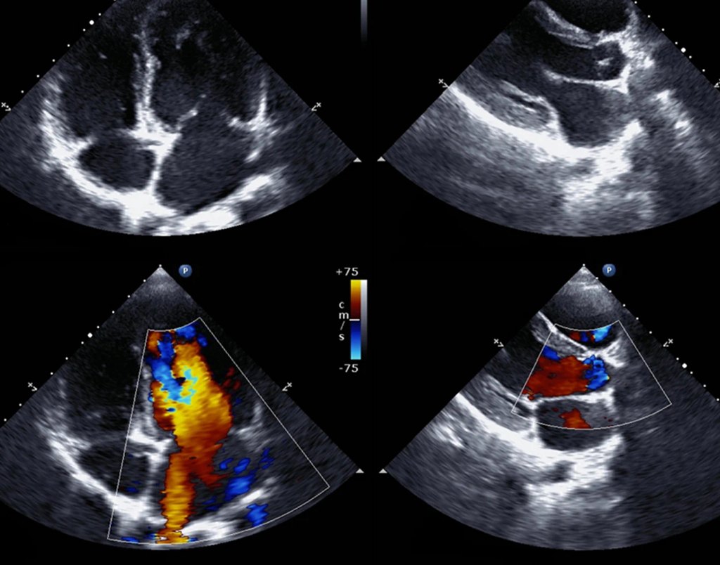
An echocardiogram (“echo”) is an ultrasound scan of the heart. It uses the same kind of high-frequency sound waves as a pregnancy scan to produce moving pictures of your heart as it beats.
An ECG tells us about the heart’s electrical activity. An echocardiogram shows us the heart itself: its chambers, muscle and valves, moving in real time. If you’ve been told you have a heart murmur, have an abnormal ECG, or have symptoms like breathlessness or ankle swelling, an echo is often the test that gives a clear answer.
This guide covers what an echo can and can’t show and whether it’s the right test for you. It also explains how our service works: the scan is done and reported by a consultant cardiologist, not a technician whose images are reviewed later.
What is an echocardiogram?
An echocardiogram (“echo”) is an ultrasound scan of the heart. It uses the same kind of high-frequency sound waves as a pregnancy scan to produce moving pictures of your heart as it beats.
It’s non-invasive and painless, and it uses no X-rays or radiation, so it can safely be repeated whenever it’s clinically useful.
At OneMedicine, scans are performed on a Philips iE33 cardiac ultrasound system with Doppler and 3D imaging.
What an echo can show
Pumping function. How strongly the left and right sides of the heart contract, usually expressed as the ejection fraction.
Chamber size. Enlargement of the atria or ventricles, which can occur with high blood pressure, valve disease, heart failure and atrial fibrillation.
Heart muscle thickness. This matters in long-standing high blood pressure and suspected cardiomyopathy.
The heart valves. Whether any of the four valves are narrowed (stenosis) or leaking (regurgitation), and how severely.
Blood flow. Doppler measures the speed and direction of blood through the heart, which helps grade valve problems and estimate pressures.
The pericardium. The sac around the heart, including any fluid collecting in it.
The aortic root and some structural or congenital abnormalities, such as holes between chambers.
What an echo doesn’t show
A standard resting echo does not look at the coronary arteries directly, so a normal echo does not rule out narrowed arteries. If your main symptom is chest pain or tightness on exertion, a different test may be more useful. See Which heart test is right for me? below, or book a consultation first.
Common reasons people book a private echo
A heart murmur heard by a GP, dentist, insurance medical or at a sports check.
An abnormal ECG, for example signs of a thickened heart muscle or an enlarged chamber.
Breathlessness, reduced exercise tolerance or ankle swelling, where heart failure needs to be ruled out or confirmed.
Palpitations or atrial fibrillation. An echo doesn’t diagnose the rhythm itself, but it shows whether there’s a structural problem behind it.
Long-standing or hard-to-control high blood pressure.
A family history of cardiomyopathy, or of sudden unexplained death in a relative under 35.
Follow-up of a known valve problem or heart condition, without waiting for the next hospital slot.
An employer, sports body, insurer or specialist has asked for one.
If you have no symptoms and no risk factors, a routine echo “just to check” is rarely needed. If you’re unsure, a short consultation will tell you whether it’s worthwhile.
Which heart test is right for me?
Test What it answers Price
Resting ECG Heart rhythm and electrical patterns £70 Book
Echocardiogram Structure, valves and pumping function £225 Book
Exercise treadmill test (exercise ECG) How your heart’s rhythm and blood pressure respond to exertion; required for DVLA Group 2 and CAA pilot medicals £250 Book
Stress echocardiogram How the heart muscle itself responds to exercise £375 Book
Cardiology consultation Not sure which test you need? Prof Khan assesses you and recommends the right investigation £185 in clinic / £125 by video Clinic · Video
An ECG and an echo often complement each other: one looks at the wiring, the other at the structure. Many patients have both.
What happens during the scan
You’ll be asked to undress to the waist. A gown is provided, and two-piece clothing makes this easier.
You lie on a couch, mostly on your left side.
Three small ECG stickers go on your chest so the images can be timed to your heartbeat.
Prof Khan applies ultrasound gel and moves a probe over your chest and just under your ribs to image the heart from several angles. You may be asked to hold your breath briefly or change position.
The scan itself usually takes 30–40 minutes, within a 45-minute appointment.
No preparation is needed. Eat, drink and take your usual medication as normal. If you’ve had an echo before, please bring or forward the report so we can compare.
Understanding your echo report
Some of the terms you may see:
Ejection fraction (EF). The percentage of blood the left ventricle pumps out with each beat. Around 55% or above is generally normal. Lower values indicate reduced pumping function.
Valve regurgitation graded trivial, mild, moderate or severe. A trivial or trace leak through the mitral or tricuspid valve is very common in healthy hearts and is usually not a concern.
Left ventricular hypertrophy (LVH). Thickening of the main pumping chamber’s wall, often linked to high blood pressure.
Diastolic function. How well the heart relaxes and fills between beats.
Because the cardiologist who performed your scan also writes the report, you get [CONFIRM: “a clear explanation on the day and…”] a report that sets out what the findings mean for you and whether any follow-up is needed.
Is an echocardiogram safe?
Yes. A standard transthoracic echo uses ultrasound only, with no radiation, no needles and no known side effects. The only discomfort is the cool gel and some light pressure from the probe.
Do I need a GP referral?
No. You can book directly online. If you already have a referral letter or previous results, bring them along.
If your symptoms are complex, especially chest pain, a cardiology consultation first makes sure you have the right test rather than just a test.
When to seek urgent help instead
An outpatient echo is not an emergency service. Call 999 or go to A&E if you have:
chest pain that is severe, lasts more than a few minutes, or comes on at rest
severe breathlessness
collapse or fainting
palpitations with chest pain, breathlessness or dizziness
Part of a complete cardiac service
The echo sits alongside the rest of our heart diagnostics at Kings Heath:
resting ECG
exercise treadmill testing
stress echocardiography
heart health blood tests
[CONFIRM: 24-hour ECG (Holter) and 24-hour blood pressure monitoring, if offered]
consultant cardiology review
Where further imaging is needed, [CONFIRM: “we can arrange CT coronary angiography or cardiac MRI”].
For full details, prices and preparation notes, see our private echocardiogram in Birmingham page.
Frequently asked questions
Who performs the scan?
Prof Jawad Khan, Consultant Cardiologist, performs and reports every echo himself.
How long does it take?
Allow 45 minutes. The scan itself takes 30–40 minutes.
When will I get my results?
24 hours
Do I need to fast or stop my medication?
No. Eat, drink and take your medication as normal.
Is there parking?
Yes, on site. We’re at 1 High Street, Kings Heath, B14 7AR, opposite Kings Heath railway station.
Can an echo detect blocked arteries?
Not directly. A resting echo shows the heart’s structure and function. For symptoms suggesting narrowed coronary arteries, a stress test or CT coronary angiography may be more appropriate. Prof Khan can advise.
Book your echocardiogram
· Consultant Cardiologist · no referral · Kings Heath, Birmingham
See available times →
Not sure it’s the right test? Book a cardiology consultation
OneMedicine is a CQC-registered clinic. This article is general information and not a substitute for personal medical advice.